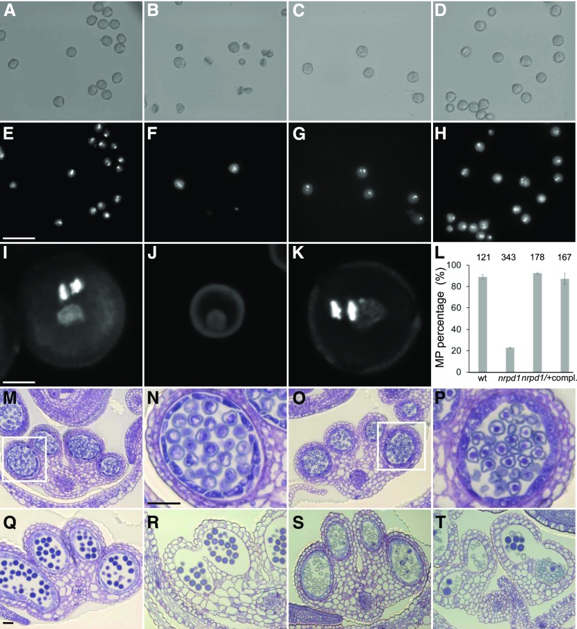
Figure 3.
Cr nrpd1 Pollen Arrest at the Microspore Stage.
(A) to (H) Bright-field ([A]–[D]) and corresponding DAPI ([E]–[H]) staining of manually dissected pollen from anthers at stage 12/13. Pollen of the wild type ([A] and [E]), Cr nrpd1 homozygotes ([B] and [F]), Cr nrpd1 heterozygotes ([C] and [G]), and a complemented line ([D] and [H]) is shown. Bar = 50 μm.
(I) to (K) Confocal images of DAPI-stained pollen of the wild type (I), Cr nrpd1 homozygotes (J), and Cr nrpd1 heterozygotes (K). Bar = 5 μm.
(L) Percentage of mature pollen (MP) in anthers dissected at stage 12/13 from the wild type (wt), Cr nrpd1 homozygotes (nrpd1), Cr nrpd1 heterozygotes (nrpd1/+), and a complemented line (compl.). Pollen was dissected from two individual plants per genotype. Error bars represent sd. Numbers of pollen grains counted in each genotype are shown above the bars.
(M) to (T) Microsporangia cross sections stained with toluidine blue at anther stages 8 ([M] and [O]), 11 ([Q] and [S]), and 12 ([R] and [T]) of the wild type ([M], [Q], and [R]) and Cr nrpd1 ([O], [S], and [T]). Insets in (M) and (O) are shown enlarged in (N) and (P), respectively. Bar for (M), (O), and (Q) to (T) = 50 μm; bar for (N) and (P) = 50 μm.

