Figure 2.
Pollen Development Was Defective in KETCH1 Loss-of-Function Mutants.
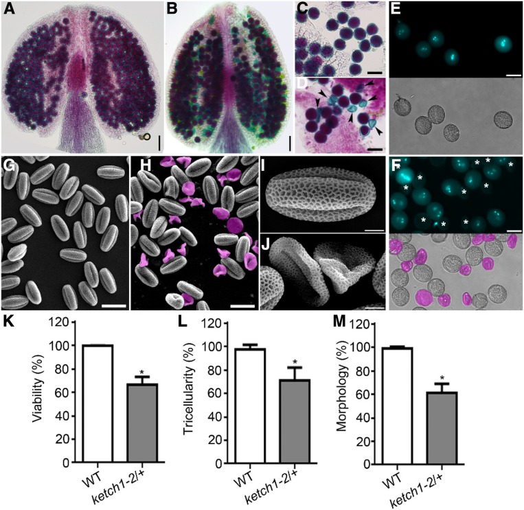
(A) to (D) Alexander staining of a representative anther (A) and (B) or mature pollen grains (C) and (D) from wild type (A and C) or ketch1-2/+ (B) and (D). Arrowheads point at aborted pollen grains.
(E) and (F) DAPI staining of mature pollen grains from wild type (E) or ketch1-2/+ (F). Bright-field (BF) images are shown at the bottom of corresponding fluorescent images. Aborted pollen grains are labeled by asterisks (in the fluorescent image) or in pink (in the BF image).
(G) to (J) Scanning electron micrographs (SEMs) of mature pollen from wild type (G) and (I) or ketch1-2/+ (H) and (J). Aborted pollen grains are highlighted in pink.
(K) to (M) Percentage of viable pollen by Alexander staining (K) of pollen with tricellular structure by DAPI staining (L) or of oval-shaped pollen by SEM (M). Results shown are means ± SD (n = 50 to 100). Asterisks indicate a significant difference of ketch1-2/+ from wild type (t test, P < 0.05). Bars = 50 µm for (A) and (B); 25 µm for (C), (D), (G), and (H); 20 µm for (E) and (F); 5 µm for (I) and (J).

