Figure 6.
KETCH1 Is Expressed in Reproductive Cells.
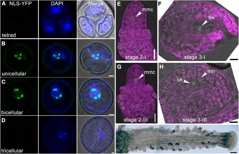
(A) to (H) CLSM of ProKETCH1:NLS-YFP in developing microspores at tetrad (A), unicellular (B), bicellular (C), tricellular microspores (D), and ovules at stage 2-I (E), stage 2-III (G), stage 3-I (F), and stage 3-III ovules (H). Dotted lines in (A) illustrate vegetative nuclei. Images in (E) to (H) are merges of the RFP (for lysotracker red staining, in magenta), YFP (for NLS-YFP, green), and transmission channels.
(I) Histochemical GUS staining of a mature ProKETCH1:GUS pistil. cn, Chalazal nucleus; fm, functional megaspore; mmc, megaspore mother cell; mn, micropylar nucleus. Two overlapping high-magnification images were taken for one pistil. The images were then overlaid with Photoshop (Adobe) to show the whole pistil. Bars = 2 µm for (A) to (D), 10 µm for (E) to (H), 100 µm for (I).

