Figure 5.
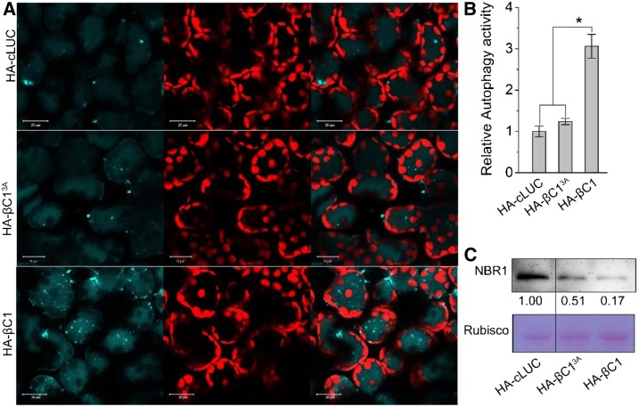
Viral βC1, but not βC13A, Is Able to Activate Autophagy.
(A) Representative confocal microscopy images of dynamic autophagic activity revealed by the specific autophagy marker CFP-NbATG8f in plants infiltrated with HA-cLUC, HA-βC13A, or HA-βC1. Autophagosomes and autophagic bodies are revealed as CFP-positive puncta in mesophyll cells. CFP-NbATG8f fusion proteins are in cyan, and chloroplasts are in red. Bars = 20 mm.
(B) Quantification of the CFP-NbATG8f-labeled autophagic puncta/cell from (A). More than 500 mesophyll cells for each treatment were used for the quantification. Relative autophagic activity in HA-cLUC-infiltrated plants was normalized to control plants, which was set to 1.0. Values represent means ± se from three independent experiments; t test used for analyses, P ≤ 0.05 (*).
(C) Immunoblot assays showed that NbJoka2 protein level was reduced due to the increased autophagy flux in HA-βC1 plants. NbJoka2 was detected with anti-NBR1 polyclonal antibody.

