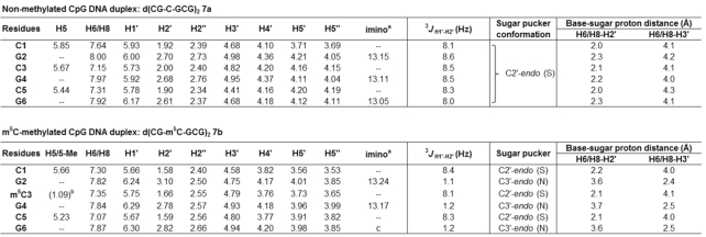
Table 3.
1H NMR chemical shifts δH (ppm) for CpG DNA duplex 7a (top) and its m5C-methylated counterpart 7b (bottom) observed at 25°C in D2O with WATERGATE suppression. The representative 2D NOESY spectrum is given in Supplementary Figure S3. For calculation of sugar-base proton distances, see Supplementary Data

|
aObserved at 10°C in 9:1 buffer/D2O solvent mix. The buffer used was 10 mM sodium phosphate buffer (pH 7.4) containing 150 mM NaCl and 20 mM MgCl2.
bChemical shift for 5-methyl group on m5C.
cNot observed even at 4°C.
