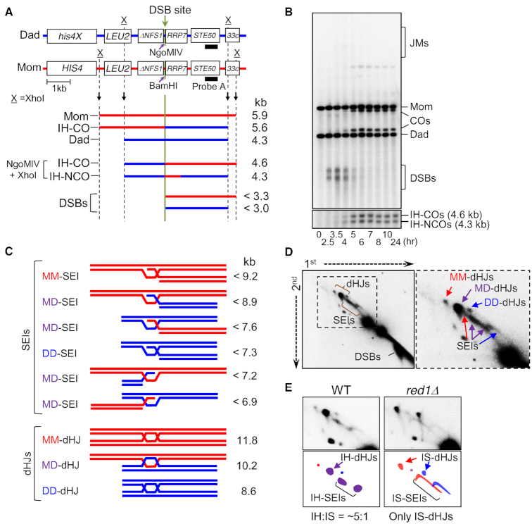
Figure 2.
System for physical analysis of DNA events of meiotic recombination. (A) Physical map of the HIS4LEU2 locus showing the DSB sites, enzyme restriction sites and the probe position for Southern blot hybridization. For physical analysis of recombination, DNA species digested with XhoI are separated on 1D or 2D gel and detected by Southern hybridization with probe A (19,20,52,69). (B) Representative image of 1D Southern analysis of WT. JMs, joint molecules; COs, crossovers; DSBs, double-strand breaks; IH-COs, interhomolog crossovers; IH-NCOs, interhomolog NCOs. (C) Diagrams of JMs, SEIs and dHJs. MM, mom–mom intersister; DD, dad–dad intersister; MD, mom–dad interhomolog. (D) 2D gels displaying meiotic recombination intermediates. Mom–mom IS, dad–dad IS and mom–dad IH species in red, blue and purple, respectively. (E) SEIs/dHJs from WT and red1Δ visualized with probe A. IS-SEI signals are spread out over a larger area due to the fact that the DSBs that are contained within IS-SEIs are hyperresected while IH-SEIs are identified at appropriate positions (19,20).

