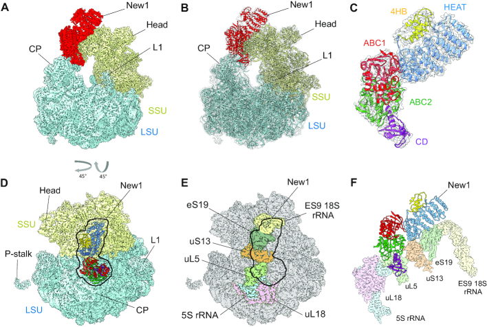
Figure 3.
The New1 interaction with the 80S Ribosome in Saccharomyces cerevisiae. (A and B) Cryo-EM reconstruction of the New1–80S complex with (A) segmented densities for New1 (red), SSU (yellow) and LSU (cyan) and (B) transparent multibody refined cryo-EM map with fitted molecular models for New1 (red), SSU (yellow) and LSU (cyan). CP, central protuberance. (C) Isolated cryo-EM map density (transparent grey) for New1 from (B) with New1 model colored according to its domain architecture, HEAT (blue), 4HB (yellow), ABC1 (red), ABC2 (green) and CD (magenta). (D) Top view of the New1 (coloured by domain as in (C)) bound to the ribosome with LSU (cyan) and SSU (yellow). (E) Outline of New1 binding site on the 80S ribosome (grey) with ribosomal components that interact with New1 colored. (F) Interaction environment of New1 (coloured by domain as in (C)) on the ribosome, with ribosomal components colored as in (E).

