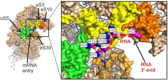
Figure 5.
The proposed binding site for unstructured RNAs near the mRNA entry site of the 40S ribosomal subunit. Solvent side view of the 40S subunit (PDB ID: 4V6X (23)). Ribosomal proteins are shown in the ‘surface’ view. The positively charged amino acid residues of the ribosomal proteins uS3 (yellow), uS5 (green) and eS30 (light orange), which could form a binding pocket for unstructured RNAs, are shown in dark blue and labeled with white rectangles. The predicted location of an RNA 11-mer is shown with a red dashed line. The residue K62 in uS3 is colored by magenta. Ribosomal protein eS10 is shown in orange.

