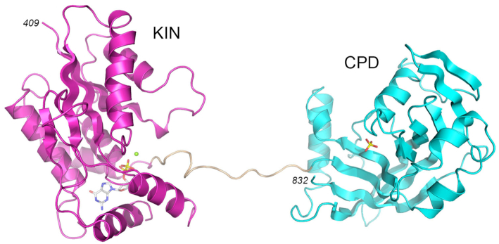
Figure 1.
Structure of Candida Trl1 KIN-CPD. The tertiary structure is shown as a cartoon model with the KIN domain in magenta, CPD domain in cyan, and the interdomain linker in beige. GDP and magnesium in the KIN active site are depicted as a stick model and a green sphere, respectively. Phosphate anion in the CPD active site is shown as a stick model.

