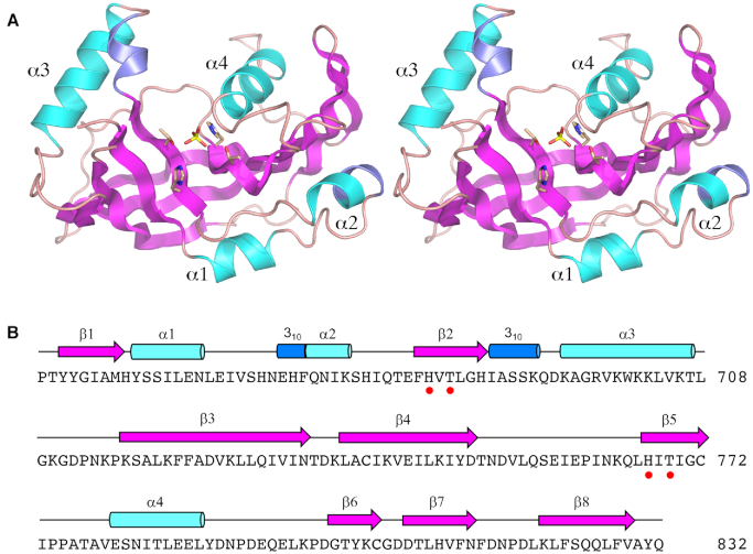
Figure 2.
Structure of the CPD domain. (A) Stereo view of the CPD tertiary structure, depicted as a cartoon model with magenta β strands, cyan α helices (numbered sequentially), and blue 310 helices. The phosphate anion in the active site and the histidine and threonine side chains that coordinate the phosphate are rendered as stick models. (B) Secondary structure elements (colored as in panel A) are displayed above the CPD primary structure. The histidine and threonine residues in the signature HxT motifs are denoted by red dots.

