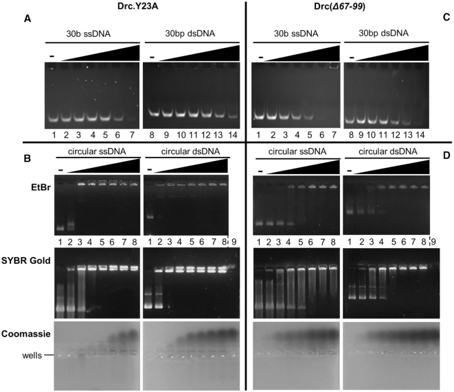
Figure 6.
(A) EMSA on native acrylamide gel with 100 nM of 5′-6-FAM-labeled ssDNA oligonucleotides (lane 1–7) or dsDNA (lane 8–14), each with increasing concentrations of Drc.Y23A (left to right: 0, 0.5, 1, 1.5, 5, 20, 50 μM). (B) EMSAs on agarose gel with 57 nM phiX174 virion DNA (left) or with its double-stranded RFI form (right). Drc.Y23A was used in increasing concentrations (lane 1 to 8; 0, 3, 6, 9, 12, 15, 18, 21 μM) for both conditions. Gels were either stained with Ethidium Bromide (top), SYBR Gold (middle) or Coomassie (bottom). A control with only 21 μM Drc, without DNA was loaded in lane 9 for SYBR Gold and Coomassie stains. (C, D) Identical to A and B, respectively, but with mutant Drc(Δ67–99). Gels are representative for two replicates.

