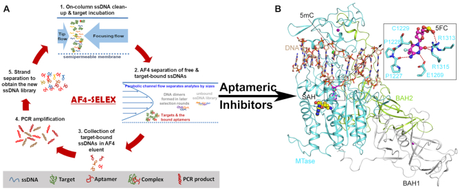
Figure 1.
(A) Scheme of AF4-SELEX. (B) Ribbon representation of mouse DNMT1 bound to CpG DNA containing a hemi methylated CpG site, in which the target cytosine was replaced by a 5-fluorocytosine (5fC) (PDB 4DA4). The DNA, BAH1, BAH2 and MTase domains are colored in wheat, grey, limon and aquamarine, respectively. The flipped-out 5fC in the catalytic site of DNMT1 is shown in the expanded view. The hydrogen bonds are shown as dashed lines. The Zinc ions and the methyl group on the 5mC are shown in purple and green spheres. The bound-cofactor analog, S-Adenosyl-L-homocysteine (SAH), is shown in sphere representation.

