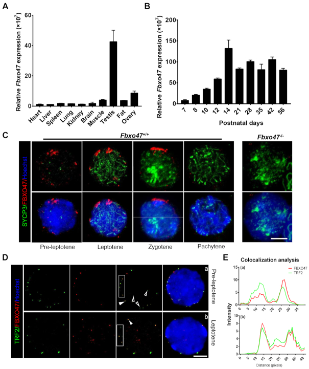
Figure 1.
Distribution pattern of FBXO47 in the testis. (A) Real-time q-PCR for Fbxo47 in various mouse tissues with 18S rRNA as a control. Error bars, SEM (n = 3). (B) Real-time q-PCR of testis samples at various postnatal days with 18S rRNA as a control. Error bars, SEM (n = 3). (C) Orthogonal z-stack projections of spermatocytes stained with the indicated antibodies. Testis cells in suspension were prepared with a mild hypotonic treatment and then fixed in Triton X-100 (39). Bars, 5 μm. (D) Nuclear colocalization of TRF2 and FBXO47 (nuclear equator). TRF2 co-localizes with FBXO47 at the INM. Arrowheads mark TRF2 located at the nuclear periphery. Hollow arrowheads mark TRF2 at the nuclear internal domain. Asterisks indicate FBXO47 at the nuclear internal domain. Bars, 5 μm. (E) Quantitative co-localization analysis of TRF2 and FBXO47 of the cell shown in panel (D).

