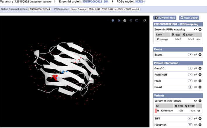
Figure 2.
The PDB model 5XRG (linked to ENSP00000221804) is displayed using LiteMol as a Richardson diagram in the central panel. rs1425150829 has been flagged in red at position 128 (ARG) occupying the end of a β strand and shows proximity to a ligand in the 3D structure, suggesting possible disruption. Additional annotation such as exons, protein domains and other variants can be turned on and off by clicking on the associated eye icon on the right hand-side of the visualization.

