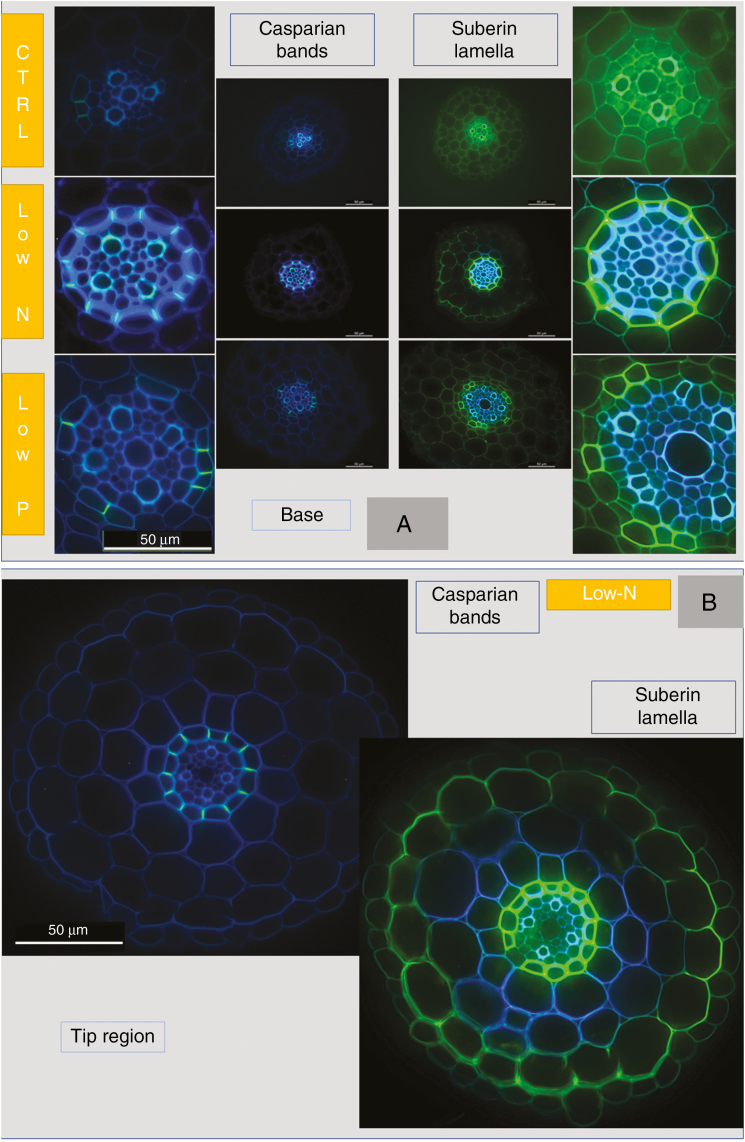
Fig. 10.
Lateral root anatomy of 14- to 18-d-old barley plants. Plants were grown on complete nutrient solution (CTRL, control) or on nutrient solution containing only 3.33 % of the nitrogen (low-N) or 2.5 % of the phosphate (low-P) of the control solution. Sections were made (A) 1–2 mm from the base of lateral roots, where they emerge from the main root axis. (B) Image taken at the tip region of lateral root of a low-N plant, showing an exodermis-like appearance of suberized structure (intense yellow signal) at the boundary between root epidermis and outermost cortical cell layer (picture on right), in addition to endodermal suberin lamella (intense yellow signal). Casparian bands (bright yellow–blueish signal), which are typical for an exodermis, could only be seen in the endodermis (left image). Scale bar = 50 µm.

