Fig. 12.
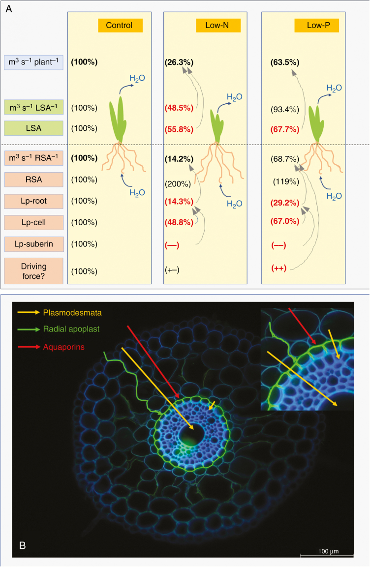
(A) Schematic of changes that affect water flow in 14- to 18-d-old barley plants in response to a low supply of N and P. The boxes in the left panel represent (from top to bottom) transpirational water flow rate per plant (m3 s−1 plant−1); transpirational water flow per leaf surface area (m3 s−1 LSA−1); leaf surface area (LSA); transpirational water flow per root surface area (m3 s−1 RSA−1); root surface area (RSA); root hydraulic conductivity (Lp-root); cell hydraulic conductivity (Lp-cell) at 15–35 mm from the tip of the main axis of seminal roots; qualitative assessment of the hydraulic conductivity of suberin lamella (Lp-suberin; 100 %, control level; –, less conductance); and driving force for radial root water uptake in a transpiring plant (Driving force?). The driving force was not measured and it can only be speculated (‘?’) whether and how (+−, no change; ++, increase) it changed in response to nutritional treatment. Values in low-N and low-P plants are expressed as percentage of the control value (set to 100 %). Arrows indicate the contributions of certain processes to some of the quantities analysed. For example, in the low-N condition reductions in transpirational water loss per LSA and also LSA contributed, together, to the reduction in the rate of plant transpirational water loss. In the same plants, the reduction in root Lp could explain, on its own, the reduction in transpirational water flow (and associated water uptake) per root surface area. (B) Flow paths for water across the root cylinder of plants, such as low-N and low-P plants, which have the endodermis completely covered with suberin lamella in the tangential walls and have Casparian bands in all radial walls. Three flow paths are distinguished: a purely apoplastic path along the radial walls, which is slowed down/blocked by Casparian bands; a path involving transport through AQPs, which may proceed up to the innermost cortex cell layer and is then slowed down/blocked by suberin lamella; and a symplasmic flow path involving transport through plasmodesmata, bypassing suberin lamella. The small picture at the top right shows part of the main picture at twice the magnification.

