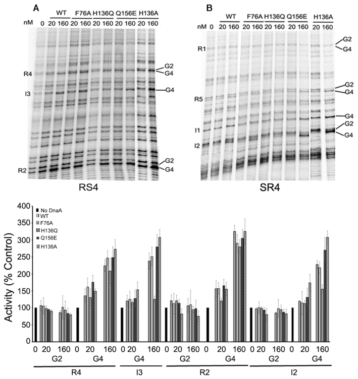
Figure 4.
DnaA(H136Q) fails to bind with low-affinity sites present within oriC. In vitro dimethyl sulfate footprint analysis was performed with WT and mutant DnaA proteins (20 nM and 160 nM) in the presence of 0.5 mM ATP to assess their occupation of low and high-affinity sites present within oriC. (Top) Two primers (A) RS4 and (B) SR4 were used to carry out primer extension to monitor changes in intensities of G2 and G4 guanines present within DnaA recognition sites. (Bottom). The band intensities of modified guanines present at R2 and R4 (representing high-affinity sites), and I2 and I3 (representing low-affinity sites) were plotted relative to respective intensities within the no-protein lanes.

