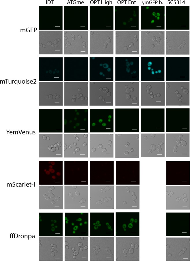
Figure 2.
Images were taken with a Fluoview 1000 confocal microscope, using the appropriate excitation and emission filters for each fluorescent protein. Cells were grown overnight on low fluorescent medium, before bringing to an optical density at 600 nm (OD600) of 0.2 in fresh Low Fluorescent medium and grown to the exponential phase (4–5 hrs) before measurement. For each codon variant one representative strain is represented. Laser intensity is kept constant between codon variants of the same fluorescent protein and also between γmGFP and ffDronpa.

