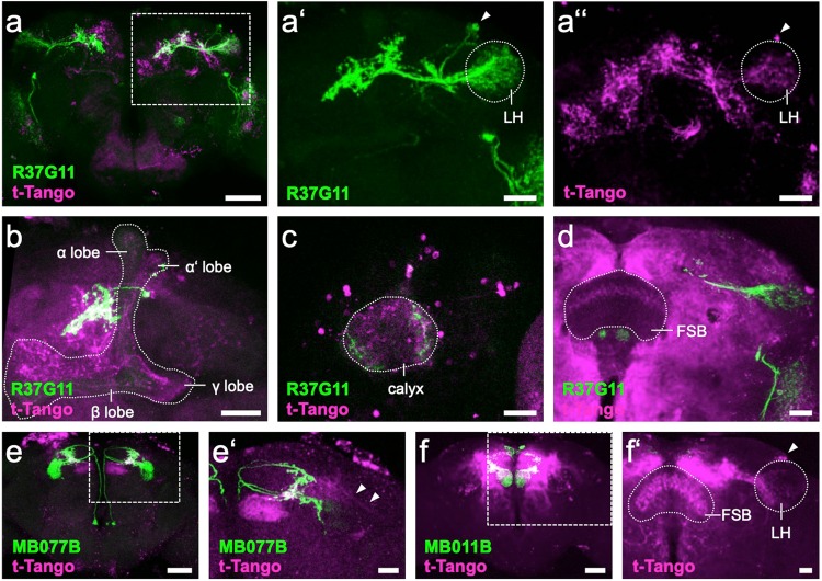
Figure 7.
trans-Tango downstream targets of PD2a1/b1, MBON- 𝛾2α’1 and MBON- β’2mp neurons. (a) R37G11-GAL4-driven myrGFP (green) labels 6-7 cells in the LH, whereas the trans-Tango signal (magenta) appears to be strongest in overlapping neuropil regions and in random sparse KC subsets in the MB (boxed area enlarged in a’ and a”). (a’) R37G11-GAL4-driven myrGFP (green) labels 6-7 cells (arrowhead) in the LH (stippled). (a”) R37G11-GAL4-driven trans-Tango signal (magenta) marks cells (arrowhead) in the LH (stippled). (b) Confocal stack of MB (stippled), highlighting the PD2a1/b1 neurites in proximity to the vertical lobe and a trans-Tango signal in all sublobes, i.e., αβ, αβ’, and 𝛾. (c) Confocal stack of MB calyx (stippled) showing innervation by R37G11-GAL4-driven GFP (green) and trans-Tango signal in both KC somata and the calyx neuropil. (d) Confocal stack of left hemibrain highlighting R37G11-GAL4-driven myrGFP (green) and postsynaptic trans-Tango signal in layer 6 of the FSB (stippled). (e) MB077B-GAL4-driven myrGFP (green) labels 3-4 MBON-𝛾2α’1 neurons, whereas a fairly restricted putative downstream trans-Tango signal (magenta) most prominently labels MBON- β’2mp (boxed area enlarged in e’). (e’) Confocal stack of left hemibrain highlighting MB077B-GAL4-driven mCD8::GFP (green) and postsynaptic trans-Tango signal (magenta) in β’2mp of the MB and in putative LHON neurites (arrowheads). (f) MB011B-GAL4-driven myrGFP (green) labels MBON- 𝛾5β2α and MBON- β’2mp, whereas the putative downstream trans-Tango signal (magenta) labels several different cell types (boxed area enlarged in f’). (f’) Confocal stack of right hemisphere highlighting the postsynaptic signal of MBON- β’2mp in PD2 LHONs (arrowhead) in the LH (stippled) and in layer 4 & 5 of the FSB (stippled). (a–f’): Confocal stack views of 1-2 µm-thick optical section data. Scale bars: a,e,f: 50 µm; a’,a”,b-d,e’,f’: 20 µm.

