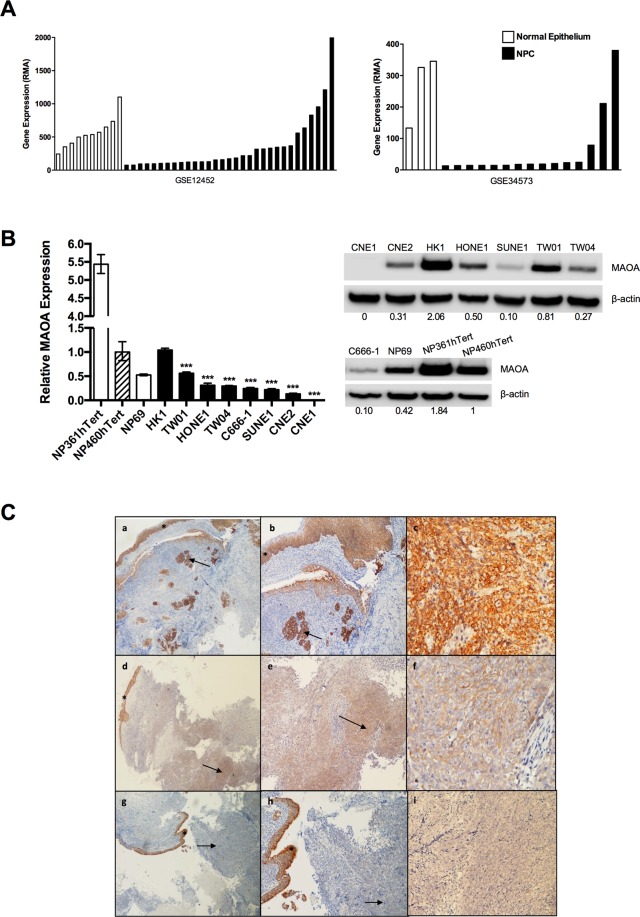
Figure 1.
MAOA is down-regulated in NPC. (A) Two microarray datasets (GSE12452 and GSE34573) showed that MAOA mRNA was significantly down-regulated in micro-dissected NPC cells compared with normal epithelium (p < 0.01). (B) Compared to NP460hTert, MAOA mRNA and protein levels were decreased in seven (C666-1, CNE1, CNE2, HONE1, SUNE1, TW01 and TW04) out of eight NPC cell lines examined. Densitometric data are expressed as the relative density (normalized to β-actin). Full-length blots are presented in Supplementary Fig. S1. ***p < 0.001. (C) Immunohistochemical analysis of MAOA expression in primary NPC. (a,b): A case demonstrating positive MAOA staining. The adjacent normal epithelium (asterisk) and underlying islands of carcinoma (arrow) are both strongly positive; (c) High-power view of area highlighted by arrow in b. (d,e): A case demonstrating moderately positive MAOA staining of carcinoma (arrow) beneath the overlying epithelium (asterisk) that is strongly positive; (f) High-power view of area highlighted by arrow in e. (g,h): A case demonstrating negative MAOA staining. The adjacent normal epithelium (asterisk) is strongly positive whilst there is no staining in the carcinoma (arrow); (i) High-power view of area highlighted by arrow in h. Original magnification (a,d,g) ×40; (b,e,h) ×100; (c,f,i) ×400.

