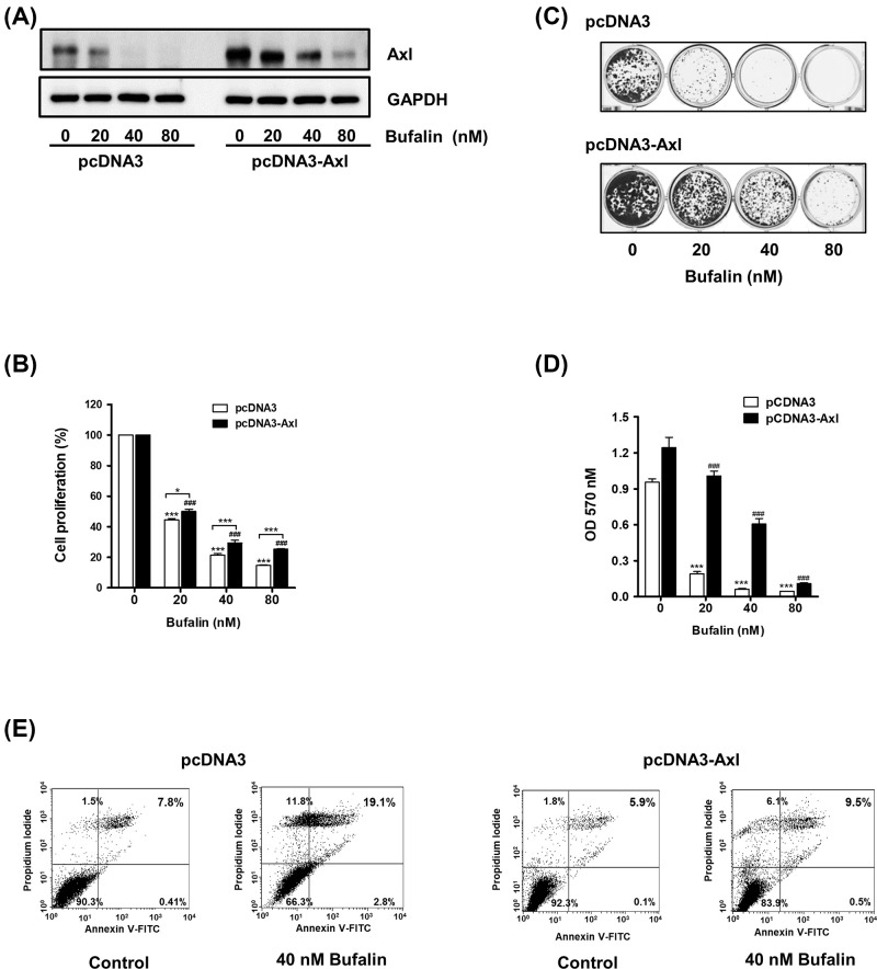
Figure 3. The inhibitory effects of bufalin on cell proliferation, colony formation, and induction of apoptosis are attenuated by Axl overexpression.
(A) H460/pcDNA3 or H460/pcDNA3-Axl cells (3 × 10⁵ cells/ 60-mm dish) were exposed to 20, 40, or 80 nM bufalin for 24 h, and Western blot analysis was conducted to determine Axl protein levels. (B) Cells (2 × 10³ cells/ 96 well) were treated with the indicated concentrations of bufalin for 24 h, and cell viability were measured using CCK-8. (***P<0.001 (H460/pcDNA3), ###P<0.001 (H460/pcDNA3-Axl), vs untreated group, *P<0.05, ***P<0.0001, H460/pcDNA3 vs H460/pcDNA3-Axl). (C) Cells (2 × 10³ cells/24 well) were treated with 20, 40, or 80 nM bufalin for 24 h, washed with PBS, and then allowed to grow for the next 7 to 10 days. The colonies were visualized by Crystal Violet staining. (D) For quantification of colony formation assay, colonies were destained and the absorbance at 570 nm was then measured. (***P<0.001, vs untreated group). (E) To detect apoptotic cells, cells were treated with 40 nM bufalin for 24 h, and the percentages of Annexin V and/or PI-stained cells were calculated.

