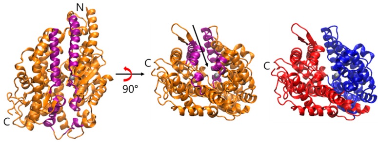
Figure 1.
The overview of the structure of C domain of sACE (PDB ID: 4APH). The ribbon representation of sACE shows the secondary structure and the two lips (purple colored) of the mouth. N and C indicate the N- and C-terminus of the enzyme, respectively. Zinc ion is shown as a gray sphere. The rightmost panel shows two subdomains that form two sides of the active site in the cleft, and the subdomain I (residues 40–122, 297–437, 551–583) and II (residues 123–296, 438–550, 584–625) are colored by blue and red, respectively. The arrow indicates the active site near the zinc ion and the putative binding pathway of ligands. The first lip (residues 73–100, 297–304, 348–354, 370–379) belongs to subdomain I, and the second (109–131, 143–156, 267–276) belongs to subdomain II.

