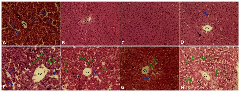
Figure 1.
Hematoxylin and eosin-stained liver slices from the treated pig. (A) Pigs fed a normal diet with 0 mg/kg Se; (B) pigs fed a normal diet with 0.3 mg/kg Se; (C) pigs fed a normal diet with 1.0 mg/kg Se; (D) pigs fed a normal diet with 3.0 mg/kg Se; (E) pigs fed a high-fat diet with 0 mg/kg Se; (F) pigs fed a high-fat diet with 0.3 mg/kg Se; (G) pigs fed a high-fat diet with 1.0 mg/kg Se; (H) pigs fed a high-fat diet with 3.0 mg/kg Se. The blue arrow indicates the sinusoids enlarged between the plates of the hepatocytes. The black arrows indicate hepatic cell necrosis. The green arrow indicates a micro- and macrovesicular fatty change. CV: central vein. (H.E. × 400).

