Figure 9.
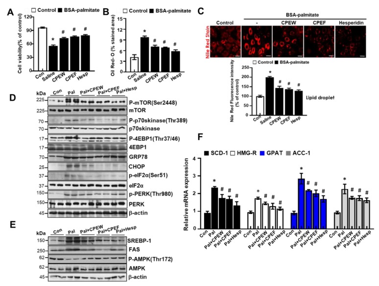
The major components of citrus peel regulate palmitate-induced steatosis in AML cells. (A) AML12 cells were treated with 100 μg/mL CPEW, 100μg/mL CPEF or 50 μM hesperidin or 24 h for the indicated periods, and cytotoxicity was determined using the MTT assay. (B) Oil-Red-O stain for lipid content staining was measured in palmitate-treated cells. (C) Nile Red stain for lipid content staining was measured in palmitate-treated cells. AML12 cells were treated with 100 μg/mL CPEW, 100 μg/mL CPEF, or 50 μM hesperidin for indicated periods, and the expression of proteins of interest was determined by western blotting. (D) Immunoblotting was performed using antibodies against p-mTOR, mTOR, p-p70S6K, p70S6K, GRP78, CHOP, p-PERK, PERK, p-eIF2α, eIF2α, and β-actin. (E) Immunoblotting was performed using antibodies against SREBP-1, FAS, p-AMPK, AMPK and β-actin. (F) mRNA expression levels including stearoyl-CoA desaturase-1 (SCD-1), 3-hydroxy-3-methylglutaryl CoA reductase (HMG-R), glycerol 3-phosphate acyltransferase (GPAT), and acetyl-coenzyme A carboxylase (ACC) was measured. Results are mean ± SEM from 3 separate experiments. (*P < 0.05 vs. control, #P < 0.05 vs. palmitate). CPEW, citrus peel extract hot-air dry; CPEF, citrus peel extract freeze-drying.*.

