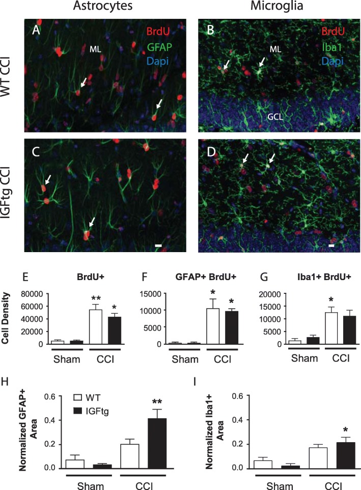
Fig. 2.
IGF1 overexpression does not potentiate posttraumatic gliogenesis in the dentate gyrus. Cells that proliferated during the first week after controlled cortical impact (CCI) detected using bromodeoxyuridine (BrdU, red) were colabelled with (a, c) the astrocyte marker, glial fibrillary acidic protein (GFAP, green) or (b, d) the microglial marker, ionized calcium binding adaptor molecule 1 (Iba1, green) in the dentate gyrus of (a, b) wildtype (WT) and (c, d) IGF1 overexpressing (IGFtg) mice at 6 weeks after injury. DAPI label is shown in blue. The scale bar represents 10 μm. GCL, granule cell layer; ML, molecular layer. (e) Density (cells/mm3) of BrdU+ cells within the ML was increased in both WT and IGFtg mice relative to their respective sham controls. Brain injury resulted in an increased density (cells/mm3) of (f) GFAP+BrdU+ cells and (g) Iba1 + BrdU+ cells within the ML relative to sham controls. IGF1 overexpression increased the relative area of (h) GFAP and (i) Iba1 immunostaining within the ML of injured mice compared to controls. Data presented as mean + SEM; n = 3 sham /genotype, n = 6–9 CCI /genotype. One-way ANOVA, followed by Bonferroni’s selected comparisons post-hoc t-tests: *p < 0.05 and **p < 0.01 compared to respective sham group

