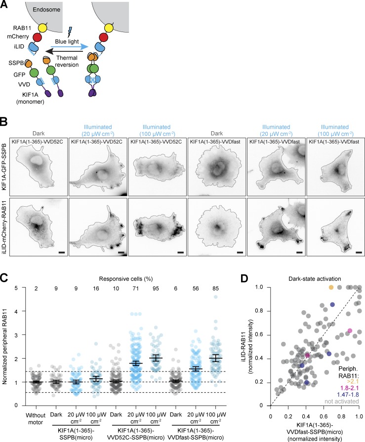
Figure 3.
Dual-layer optogenetic control limits dark-state activation of transport. (A) Assay: blue light illumination induces both dimerization of the opto-kinesin KIF1A(1–365)-VVD-GFP-SSPB(micro) and binding of the KIF1A motor to iLID-mCherry-RAB11, together stimulating plus end–directed intracellular transport of recycling endosomes into the cellular periphery. (B–D) Fixed-cell imaging (B) of indicated constructs in COS-7 cells in the dark or after illumination with low-intensity (20 µW cm−2, as in Fig. 1, B and C) or high-intensity (100 µW cm−2) blue light. Quantification in C shows the NPF of RAB11 intensity. Each dot represents one cell and bars indicate mean and 95% confidence intervals. Dotted lines represent the mean and the mean ± two times the SD of control cells. Numbers show the percentage of responsive cells for each dataset (peripheral enrichment greater than mean + two times the SD of control). Data were from at least three experiments. Graph in D shows normalized cellular expression levels of indicated constructs per cell in the dark, color coded for peripheral RAB11 intensity to indicate cells that were nonresponsive (gray) or showed dark-state activation (yellow, magenta, and blue). Scale bars are 10 µm.

