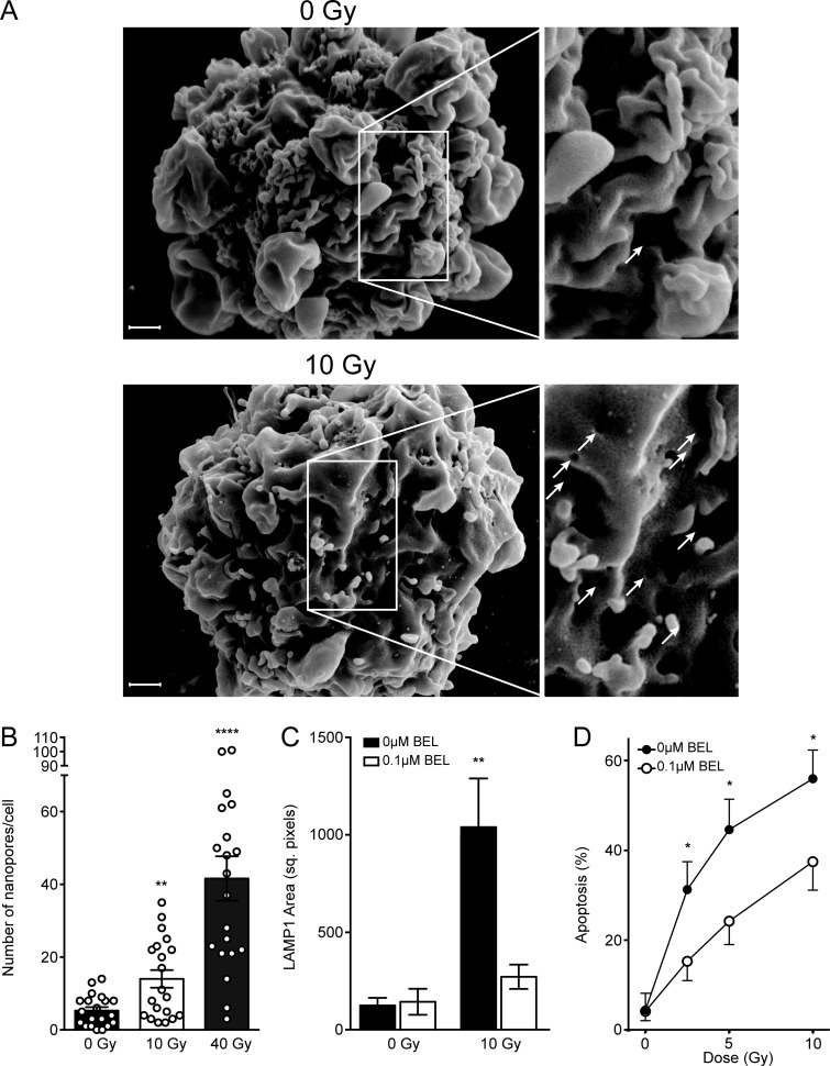
Figure S5.
Radiation induces nanopores and triggers lysosomal exocytosis-mediated apoptosis in BAECs. (A) Representative scanning electron micrographs of BAEC plasma membrane nanopores (bottom, white arrows in inset) 1 min following 10-Gy exposure. (B) Nanopore formation at 1 min is dose dependent up to 40 Gy, quantified from scanning electron micrographs. (C and D) The lysosomal exocytosis inhibitor BEL blocks LAMP1 surface staining (C) as detected by anti-LAMP1 antibody 2 min after 10 Gy and dose-dependent apoptosis (D) as measured by bisbenzimide staining 8 h after exposure. Note that as the radiation dose escalates, ASMase-independent DNA damage–mediated death becomes more pronounced, consistent with published literature (Garcia-Barros et al., 2003). Data represent mean ± SEM (A–C) and mean ± 95% confidence interval (D) and are derived from 20 cells per group (B), ≥7 cells per group (C), and ≥200 cells per group (D). **, P < 0.01; ****, P < 0.0001, two-tailed Student’s t test versus 0 Gy control (A–C) and 0 µM BEL (D). Scale bar: 1 µm. Inset magnification 2×.

