Figure 3.
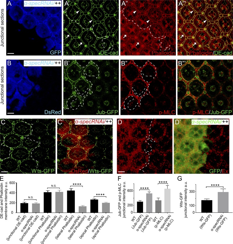
Spectrin is not required for attaching AJ-associated F-actin to cell membrane or transmission of cortical force to AJs. (A–A”’) A Triton X-100–permeabilized pupal eye disc containing GFP-positive MARCM clones with α-spec RNAi was stained for the AJ marker DE-cad and phalloidin. Note the normal DE-cad and circumferential F-actin cables associated with AJs in α-spec mutant PECs (arrowheads in A’–A”’) compared with those in the neighboring wild-type PECs (arrows in A’–A”’). Quantification of DE-cad and phalloidin intensity is shown in E. (B–B”’) A pupal eye disc containing DsRed-positive clones with α-spec RNAi was imaged for Jub-GFP and stained for p-MLC. Note the increased level of Jub-GFP at AJs (B’) and increased cortical p-MLC (B”) in α-spec mutant PECs. Quantification of Jub-GFP and p-MLC junctional intensity is shown in F. (C and C’) Similar to B–B”’ except that Wts-GFP was examined. Note the increased level of Wts-GFP in α-spec mutant PECs (C). Quantification of Wts-GFP junctional intensity is shown in G. (D and D’) A pupal eye disc containing GFP-positive MARCM clones with α-spec RNAi was stained for Ex. Note the elevated Ex level in clones with α-spec RNAi. (E–G) Quantification of mean membrane intensity of the indicated proteins in the specified membrane domains. Data are means ± SEM (n ≥ 20 cells, representative of 10 pupal eyes). ****, P < 0.0001. Scale bars, 5 µm. N.S., no significance.

