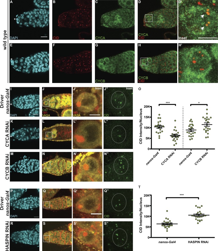
Figure 2.
CID deposition in GSCs requires CYCA, CYCB, and HASPIN. (A–H) Wild-type germaria stained for DAPI (cyan), anti-CID (red), and anti-CYCA or anti-CYCB (green). (I–N″) Confocal z-stack projection of nanos-Gal4 (I–J″), CYCA RNAi (K–L″), CYCB RNAi (M–N″) germaria, stained for DAPI (cyan), anti-VASA (yellow), anti-CID (green), and anti-1B1 (spectrosome, red). (O) Quantification of CID fluorescence intensity at centromeres per nucleus (L). (P–S″) Confocal z-stack projection of nanos-Gal4 (P–Q″) and HASPIN RNAi germaria (R–S″), stained for DAPI (cyan), anti-VASA (yellow), anti-CID (green), and anti-1B1 (spectrosome, red). (T) Quantification of CID fluorescence intensity (MGVs) at centromeres per nucleus. Data are represented as the mean ± SEM; ***, P < 0.0005; *, P < 0.05, calculated with unpaired t test with Welch’s correction. Star indicates the terminal filament and arrows indicate centromeres; 3-d-old female flies; scale bar, 10 µm; inset, 5 µm.

