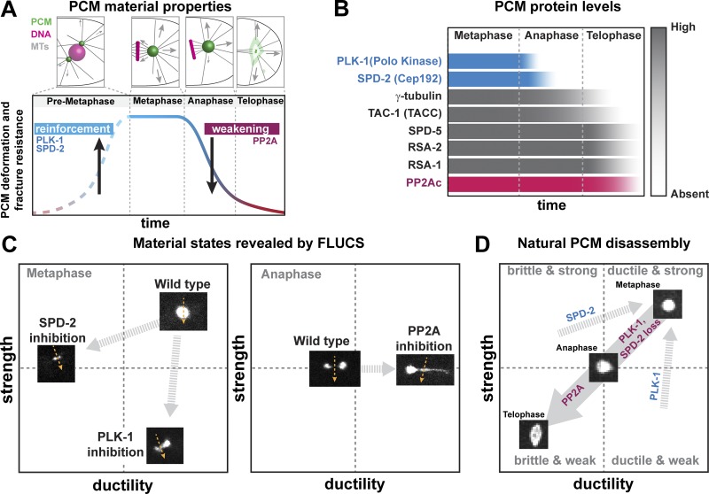
Figure 7.
The balance of PLK-1, SPD-2, and PP2A activities tunes PCM strength and ductility. (A) PCM resistance to microtubule-mediated forces peaks in metaphase during spindle assembly and then declines in anaphase and telophase, corresponding to PCM disassembly. (B) PCM-localized levels of eight different proteins during mitotic progression. In anaphase, PLK-1 levels decline first, followed by SPD-2. The catalytic subunit of PP2A phosphatase (PP2Ac), as well as the main scaffold protein SPD-5, remain at the PCM until late telophase. (C) In metaphase, FLUCS cannot fracture or deform wild-type PCM. However, FLUCS can fracture PCM in spd-2 mutant embryos (i.e., PCM is less ductile) and stretch and deform PCM in PLK-1–inhibited embryos (i.e., PCM is less strong but still ductile). In anaphase, FLUCS easily deforms and fractures wild-type PCM, while it deforms and stretches PCM in PP2A-inhibited embryos (i.e., PCM is more ductile). (D) The combination of PP2A phosphatase activity and the departure of PLK-1 and SPD-2 transitions PCM from a strong, ductile state in metaphase to a weak, brittle state in telophase. This transition enables PCM disassembly and dispersal through microtubule-mediated pulling forces.

