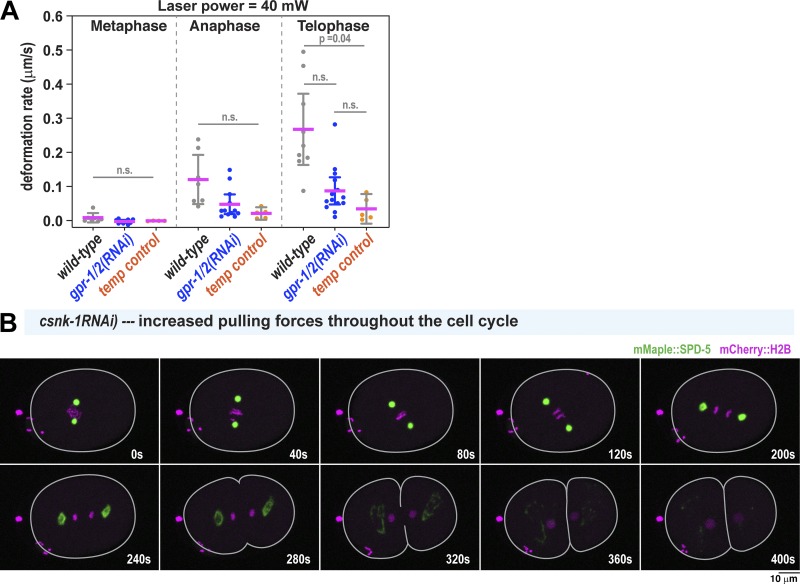
Figure S2.
Contributions of microtubule pulling forces to PCM deformation. (A) PCM deformation rates in metaphase (M), anaphase (A), and telophase (T) using high flow in wild-type and gpr-1/2(RNAi) embryos or 40-mW bidirectional laser scanning (temperature control; no flow). Data are from experiments in Figs. 1 and 2. Individual data points are plotted with mean ± 95% CI; n = 7, 7, and 9 (wild-type: metaphase, anaphase, telophase); n = 10, 12, and 13 (gpr-1/2(RNAi)); and n = 4, 5, and 5 (temperature control). P values were calculated using Brown–Forsythe and Welch ANOVAs followed by Dunnett’s T3 multiple comparisons test. n.s., not significant. (B) Time-lapse images of centrosomes in a csnk-1(RNAi) embryo, where microtubule-mediated pulling forces at the cortex are ∼1.5-fold elevated compared with wild type (Panbianco et al., 2008). PCM deformation does not occur prematurely in metaphase.

