
Figure 2.
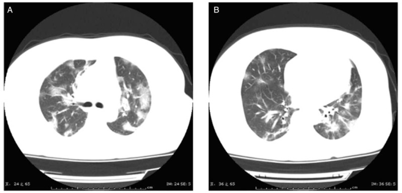
Chest CT images of a 47-year-old female patient upon admission, who had symptoms of fever for 7 days and post-exertional shortness of breath for 2 days (A and B). Transverse chest CT images showed the bilateral multiple lobular and sub-segmental areas of consolidation. CT: Computed tomography.