FIGURE 2.
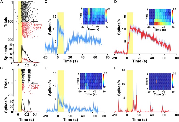
Widespread melanopsin responses are not observed in the intact retina. Raster plots and PSTHs of melanopsin-responsive unit (A) as well as a representative ON–OFF unit (B) exposed to a 1 Hz 100 ms flash (log 15 photons/cm2/s at time 0) reveal progressive loss of responses to this short duration stimulus following wash in of 100 μM DL-AP4 + 2 μM ACET starting at trial 100 (indicated in red). By trial 200, neither unit is responsive. Red PSTHs are of the last 30 stimulus presentations. (C–F) PSTHs and TBC (inserts) responses to 10/120 s stimuli of example a GCL unit (from A) classified as melanopsin-responsive from a wild type (wt) animal before (C) and after (D) pharmacological deafferentation from outer-retinal photoreceptors. PSTHs (±SEM) and TBC (inserts) responses to 10/120 s stimuli of example unit from a melanopsin knockout animal with a sustained response before (E) but not after (F) pharmacological deafferentation.

