Figure 1.
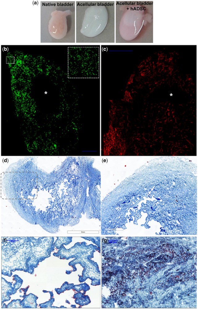
In vitro recellularization of bladder matrices with human ADSC. (a) Macroscopic view of a dissected native rat bladder (left panel), decellularized bladder after decellularization process (central panel) and decellularized bladder recellularized with labeled human ADSC (right panel); (b) confocal images of the in vitro recellularized matrix bladder with ADSC labeled with cell tracker [green (pseudo-color), left panel] and (c) phalloidin (red, right panel) and cultured for 5 DIV demonstrate a homogeneous cell distribution along the collagen-rich matrix. The inset square in (b) depicts a magnification of the indicated area. The internal lumen of the bladder is empty (*); (d and e) Masson’s trichrome staining of decellularized bladder matrix, (e) enlarged image of dotted line marked area in (d); (f and g) trichrome staining of neobladders after 5 DIV demonstrates the contrast in the collagen-rich matrix stained in blue-green and the ADSC in purple-red lined within the urothelial layers (f) and growing in clusters in the mucosa and submucosa areas (e). Scale bar, in blue: 100 μm.

