Figure 4.
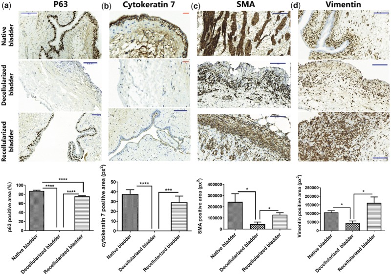
Recellularization of decellularized bladder matrices with ADSC accelerates endogenous bladder regeneration in vivo. Upper panels: (a) Representative images of p63 immunostaining (urothelial marker) and (b) cytokeratin 7 (epithelial cell marker) demonstrates a stratified transitional epithelium in the recellularized bladder matrices compatible with the native bladder. Ten days after implantation, the decellularized bladder matrices failed to demonstrate positive staining in the former urothelial layer; (c) Representative images of SMA specific staining in longitudinal and transversal orientations at the submucosa in the recellularized bladder matrices demonstrate a comparable muscle layer organization to that of native tissue. Ten days after implantation, the decellularized bladder matrices exhibit an SMA-positive population of cells lacking a defined orientation; (d) Representative images for vimentin immunostaining evidence massive mesoderm-derived cell infiltration in the recellularized bladder matrices, mostly located at the external perimeter of the recellularized neobladder. Ten days after implantation, the decellularized bladder matrices exhibit few vimentin-positive cells. Scale bars: red: 100 μm, blue: 50 μm; lower panels: graphical quantification. Data are expressed as mean ± SEM. *P < 0.05; ***P < 0.001; ****P < 0.0001.

