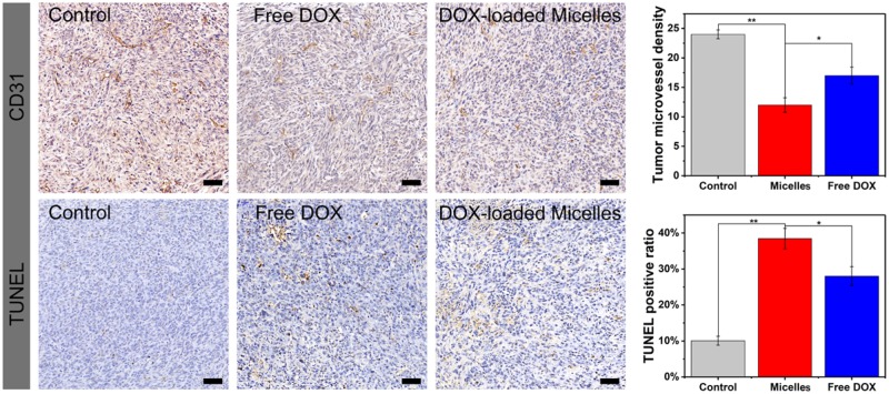
Figure 9.
CD31 and TUNEL Immunohistochemical (IHC) staining of tumor tissues treated with saline (control), free DOX and TP-PEI (DA/DOX)-PEG prodrug micelles. The brown areas demonstrated CD31-positive and TUNEL-positive staining. The scale bars were 20 μm. The capillary number was counted for CD31 (**P < 0.01, *P < 0.05). The proportion of apoptotic cells was calculated as the apoptotic indices (**P < 0.01, *P < 0.05)

