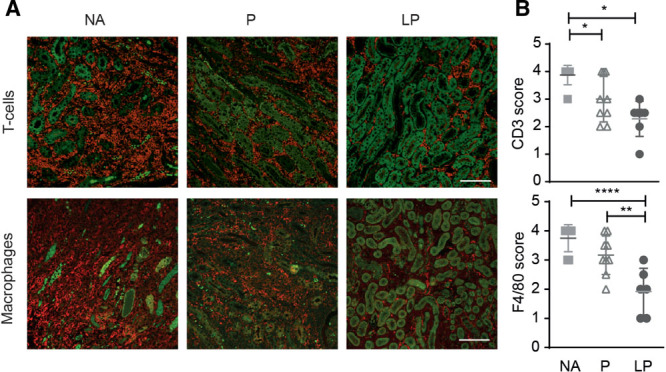
FIGURE 7.

LP treatment reduced number of F4/80+ macrophages and CD3+ T cells in the renal allografts. A, Immunohistochemistry for CD3+ T-cells (red, upper panel) and F4/80+ macrophages (red) is shown in the allograft. Dense interstitial infiltrates were present in the NA and P group and were markedly reduced in the LP-treated recipients. Green is due to the autofluorescence of the tubuli (bar: 100 µm). B, Semi-quantitative analysis confirmed that CD3+ T-cell infiltration was reduced by prednisolone and even more by LP treatment (NA; N = 8, P; N = 9, LP; N = 7). F4/80+ macrophage infiltration was reduced by LP treatment compared with NA or prednisolone (NA; N = 8, prednisolone; N = 9, LP; N = 6) (mean ± SD. *P < 0.05, **P < 0.01, ****P < 0.0001, NA = no additional treatment, P = Prednisolone, LP = liposomal prednisolone.
