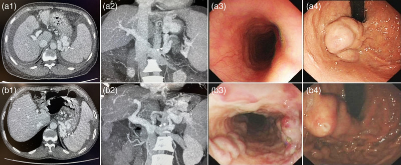
Fig. 1.

. (a) A 54-year-old female bleeding from IGV1. (a1 and a2) Enhanced CT showing solitary GV bolus (a1, white arrow) and GRS (a2, white triangles); (a3 and a4): endoscopic examination showing only GV but no esophageal varices (EV). (b) A 57-year-old male bleeding from GOV2. (b1 and b2) Enhanced CT showing GV bolus (b1, white arrow) and GRS (b2, white triangles); (b3 and b4) endoscopic examination showing both EV and GV. CT, computed tomography.
