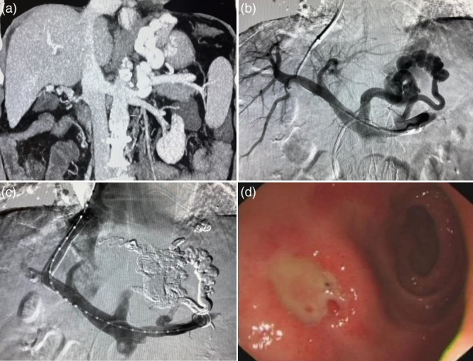
Fig. 3.

A 67-year-old male bleeding from IGV1. (a) Before the BAATO + TIPS procedure, enhanced CT showed solitary GVs bolus and GRS. (b) During the procedure, the portography showed GVs feeded of posterior gastric vein and short gastric vein; (c) post-stenting venogram showed that cyanoacrylate spreaded and embolized GVs all over. (d) After the BAATO + TIPS procedure, endoscopic examination showed the complete regression of GVs. CT, computed tomography; GRS, gastrorenal shunt.
