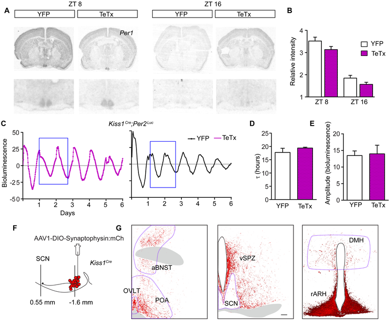
Figure 6. Kiss1ARH Neurons Send Sparse Projections to the SCN and are Not Necessary for Circadian Clock Gene Expression in the SCN.
A) Autoradiographic images of coronal brain sections radiolabeled with a Per1 probe at ZT8 or ZT16.
B) Average SCN pixel intensity in autoradiographic films. One-way ANOVA: YFP (CT8 n = 8; CT16 n = 6), TeTx (CT8 n = 8; CT16 n = 4); F(3,24) = 5.67, p < 0.0001. Bonferroni post hoc comparison at CT8 and CT16. YFP was not different from TeTx at either time point.
C) Representative traces of Per2-luc bioluminescence rendered from time-lapsed images taken every 20 min over 6 days from control and Kiss1ARH-silenced mice.
D) Average period (t) from 6 d of luminescence recordings comparing YFP-injected controls to Kiss1ARH-silenced females. Student t-test, YFP n = 3, t = 17.9 ± 1.57 h; TeTx n = 3, t = 19.4 ± 0.53 h, t(4) = 1.57, p = 0.19.
E) Average amplitude for 2 d of luminescence recording (blue box in E). Student t-test, YFP n = 4, amplitude = 26.91 ± 5.36 arbitrary units; TeTx n = 6, amplitude = 27.95 ± 12.68 arbitrary units, t(8) = 0.15, p = 0.88.
F) Schematic diagram of the targeted viral injection of a conditional synaptophysin-fused-mCherry-reporter transgene into the ARH of Kiss1cre females and the sagittal location of the ARH with respect to the SCN.
G) Kiss1ARH fiber expression throughout the mid-rostral hypothalamus, including the SCN. Tissue was immuno-stained for the mCherry reporter (A). Scale bar, 100 mm. Abbreviations: anterior bed nucleus of the stria terminalis, aBNST; organum vasculosum lamina terminalis, OVLT; pre optic area, POA; ventral subparaventricular zone, vSPZ; suprachiasmatic nucleus, SCN; dorsal medial hypothalamus, DMH; arcuate nucleus of the hypothalamus, ARH.
See also Figures S5 and S6

