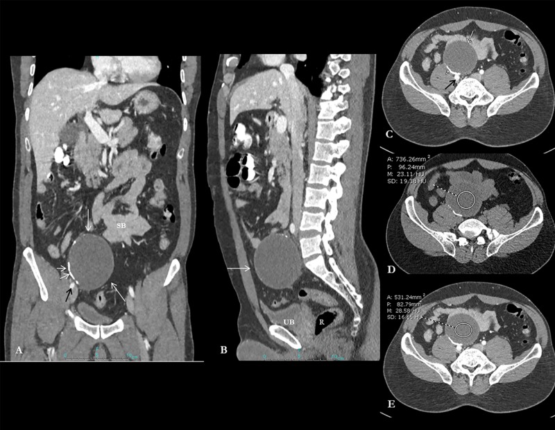
Figure 1. Composite image showing contrast-enhanced CT abdomen and pelvis coronal reformatted views.
Contrast-enhanced CT abdomen and pelvis coronal reformatted image (A) demonstrates a fairly well-circumscribed oval cystic lesion in the right hemipelvis (long white arrow). The cyst is lying anterior and separate from the right common iliac vessels (black arrow; image A and axial image C), urinary bladder (UB), rectum (R) and small bowel loops (SB) (image A, B and C). Note the presence of curvilinear hyperdense calcification (short white arrows; image A, C). Axial unenhanced image (D) and post-contrast image (E) confirm the cystic unenhanced nature of the lesion (23-28 HU).

