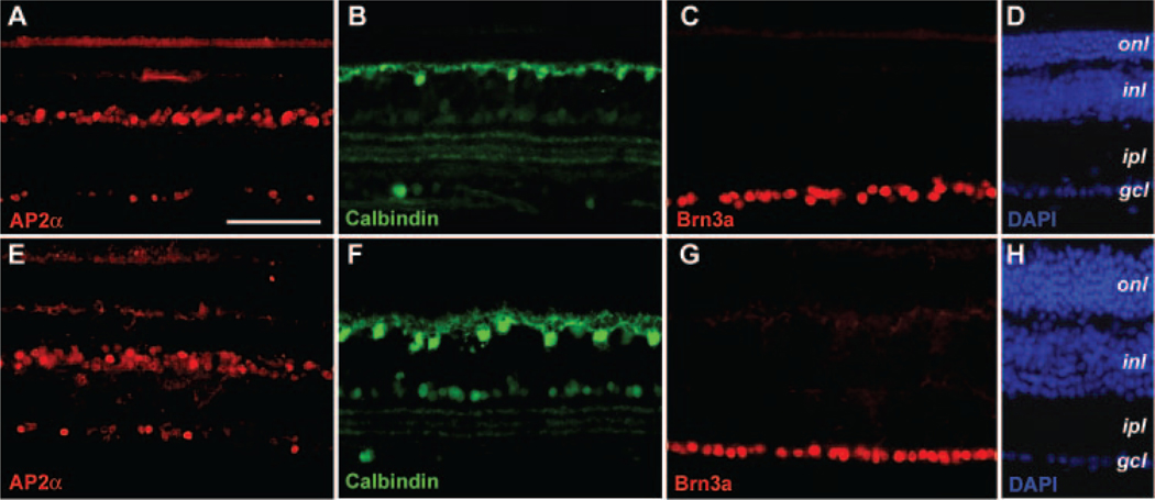
FIGURE 4.
Effects of CNTF on other retinal cell types in the rds retina. Immunofluorescent images of P90 rds retinal sections treated with rAAV-CNTF at P23 (E–H) and the untreated control retinas (A–D) labeled for amacrine cell marker AP2α (A, E), horizontal cell marker calbindin (B, F), ganglion cell marker Brn3a (C, G), and 4′,6′-diamino-2-phenylindole (DAPI; D, H) are shown. Abbreviations as in Figure 3. Scale bar, (A) 100 μm.

