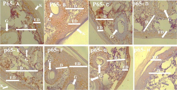
Figure 3.
Photomicrograph of the skin of the experimental animals in Groups A–H demonstrating the skin using the p65 immunohistochemistry technique (p65 A-H). More cells in the groups B, D, F, and G expressed p65 which was used as an inflammation marker.
Note: Arrows point to specific features that are denoted by letters.
Abbreviations: ED, Epidermis - (1) Stratum corneum (2) Stratum Granulosum (3) Stratum Spinosum (4) Stratum Basale; D, Dermis; G, Gland; C, Connective tissue; K, Keratin.

