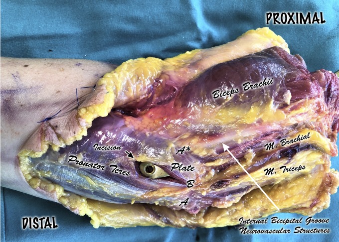
Fig. 1.
Dissection of the distal part of the right arm and elbow (semilateral view). A = apex of medial epicondyle, and A* = median nerve. Neurovascular structures run along the internal bicipital groove lying over the brachialis muscle and lateral to the pronator teres muscle belly. A to A* = the distance between the apex of the medial epicondyle and the median nerve. B = proximal tip of the incision, starting 1 cm lateral to the tip of the medial epicondyle and following its fibers distally for 2.5 cm. B to A* = the distance between the proximal tip of the muscular incision and the median nerve. A thick layer of pronator teres muscle belly was left laterally, protecting the neurovascular structures.

