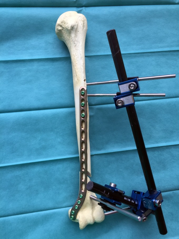
Fig. 2.

Anatomical model of a left humerus with a distal shaft fracture, used to verify the possibility of using an external fixator to reduce the fracture. Two pins were introduced distally, 1 of them into the lateral center of the capitellum and the other, just above the coronoid fossa through a small incision very close to the external border of the biceps tendon. Another 2 pins were introduced proximally in the anterolateral zone.
