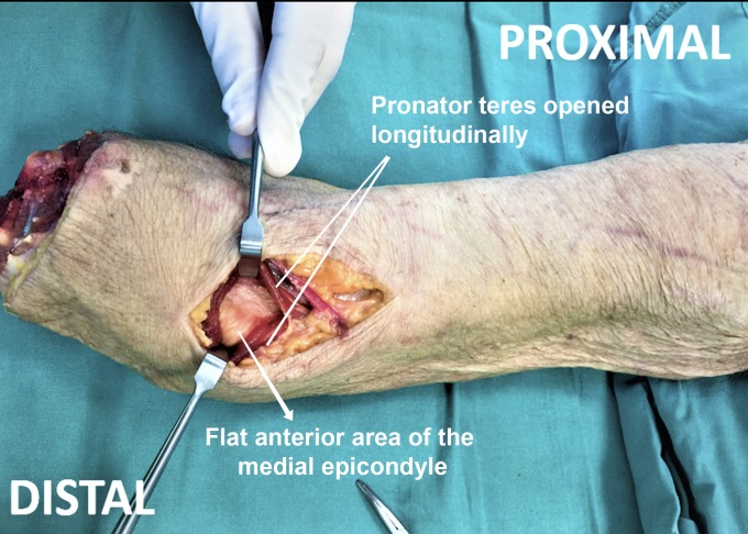
Fig. 4.
Frontal view of a right arm specimen, showing the distal approach. A 6-cm skin incision has been performed between the inner portion of the biceps tendon and the apex of the medial epicondyle. The medial antebrachial cutaneous nerve and the basilic vein have been retracted (not shown). The pronator teres muscle is then longitudinally dissected to access the anterior flat area of the medial epicondyle.

