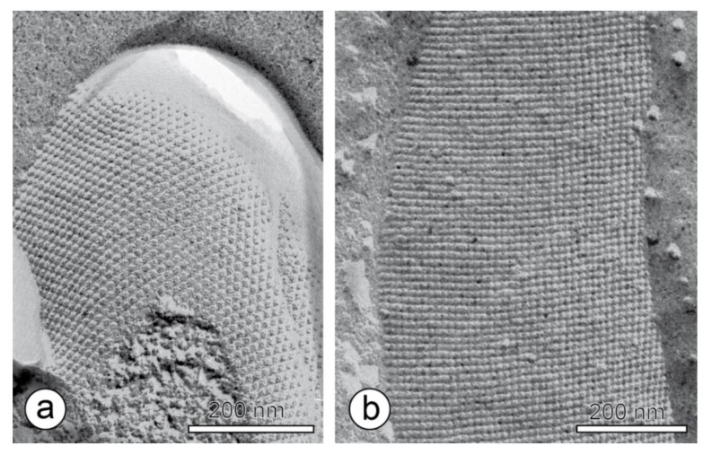
Figure 3.
Transmission electron microscopy image of a freeze-etched and metal shadowed preparation of (a) an archaeal cell (from Methanocorpusuculum sinense), and (b) a bacterial cell (from Desulfotomaculum nigrificans). Bars, 200 nm. With permission from Sleytr et al. 2014 [7] (CC BY-NC-ND 3.0).

