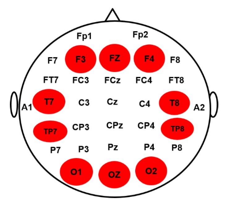
Figure 2.
The electroencephalography (EEG) channels placement in a 32-channel EEG cap according to the international 10–20 system were used for data collection. The highlighted brain regions including frontal cortex (F3, FZ, F4), occipital cortex (O1, OZ, O2), left temporal cortex (T7 TP7) and right temporal cortex (T8, TP8) were used for brain connectivity analysis.

