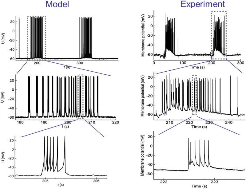
Fig 1. Single neuron activity during ictal discharges in simulation with the orignal Epileptor-2 model (left) and experiment (right).
Two ictal discharges as bursts of clustered interictal-like short bursts are seen in the membrane voltage. In the experiment, the ictal discharges were recorded in a pyramidal neuron from a rat entorhinal cortex using an in vitro 4-AP model of epileptic activity. Modified from [21].

