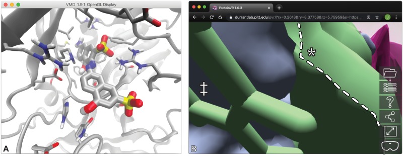
Fig 4. Two examples that show the advantages of ProteinVR use.
A) An illustration of compound V2 docked into the REL1 ATP-binding pocket, visualized using VMD. Despite the use of fog and perspective view, perceiving critical protein/ligand interactions without true stereoscopic 3D is at times challenging. B) An illustration of the space between open- and closed-pocket LARP1 surfaces, visualized using ProteinVR in non-VR mode. Prior to entering VR mode, camera-adjacent sections of the surfaces are often clipped (highlighted with a white dotted line), as is typical of non-VR programs. Clipping is easier to avoid in VR because the camera position is finely controlled by simple head movements, and VR provides a wider field of view.

